Figure 2.
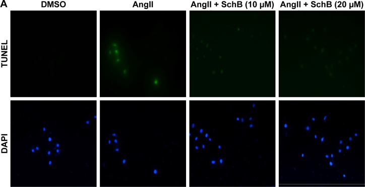
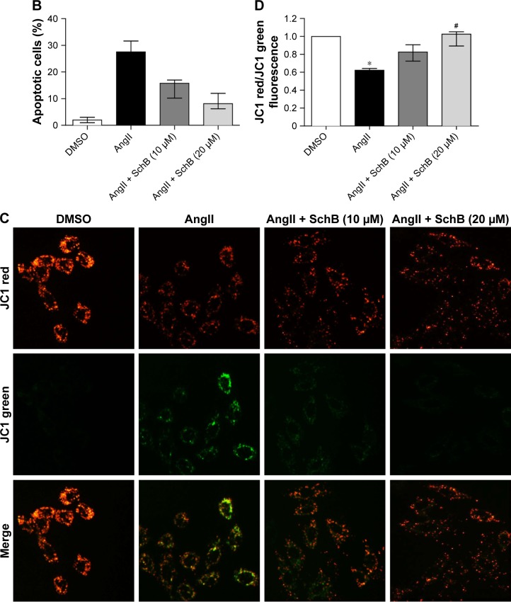
SchB inhibited AngII-induced apoptosis and MMP depolarization in RAECs.
Notes: RAECs were pretreated with SchB (10 or 20 µM) for 1 hour and then incubated with AngII (1 µM) for the times indicated. After exposure to AngII for 24 hours, the effect of SchB on AngII-induced apoptosis in RAECs was determined by TUNEL staining. Representative images for TUNEL staining are shown (A), with quantitative columns for TUNEL-positive cells (B). (C) Effect of SchB on prevention of AngII-induced mitochondrial dysfunction was assessed by JC1 staining. Increased green and reduced red fluorescence in the AngII group (24 hours’ exposure) is indicative of MMP alteration. This alteration was normalized in cells treated with SchB. (D) Quantitative analysis of the ratio of red:green fluorescence (*P<0.05 compared to DMSO; #P<0.05 compared to AngII). Magnification: 200× amplification (A); 400× amplification (C).
Abbreviations: AngII, angiotensin II; DMSO, dimethyl sulfoxide; MMP, mitochondrial membrane potential; RAECs, rat aortic endothelial cells; SchB, schisandrin B.


