Fig. 2.
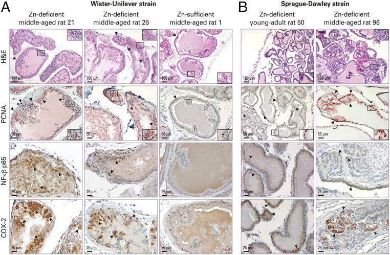
Zn-deficient middle-aged rat prostate shows increased cellular proliferation and inflammation. H&E analysis of histology and IHC analysis of PCNA, NF-κβ p65, and COX-2 protein expression were performed in FFPE prostate tissues. (A) Zn-deficient middle-aged Wistar-Unilever rat prostate vs. Zn-sufficient counterparts (n = 10 rats per dietary group). (Top Row) H&E-staining shows representative prostates from Zn-deficient middle-aged rats no. 21 and 28 displaying proliferative epithelia (arrowheads) and prostate from Zn-sufficient middle-aged rat 1 showing a thin epithelium (arrowhead) with infolding. (Scale bars: 100 μm in main panels and 50 μm in Insets.) (Second Row) In PCNA IHC, Zn-deficient middle-aged prostates show abundant PCNA-positive nuclei (red; 3-amino-9-ethylcarbazole substrate-chromogen; arrowheads); Zn-sufficient counterparts display few PCNA-positive nuclei. (Scale bars: 50 μm in main panels and 25 μm in Insets.) Additionally, Zn-deficient middle-aged prostates showed strong cytoplasmic NF-κβ p65 (Third Row) and intense COX-2 (Bottom Row) expression with typical perinuclear cytoplasmic staining; Zn-sufficient middle-aged prostates showed no NF-κβ p65 staining and occasional COX-2–positive staining (brown, DAB). (Scale bars: 25 μm.) (B) Compared with Zn-deficient young-adult rat prostate, Zn-deficient middle-aged prostate was proliferative (H&E staining, Top Row) (Scale bars: 100 μm in main panels and 50 μm in Insets) with frequent PCNA-stained nuclei (Second Row) (Scale bars: 50 μm in main panels and 50 μm in Insets), moderately strong cytoplasmic NF-κβ p65 expression (Third Row) and intense COX-2 expression (Bottom Row) (Scale bars: 100 μm).

