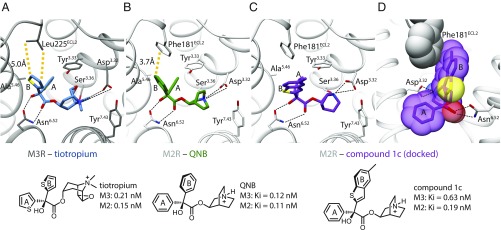
Fig. 1.
Comparison of the orthosteric binding sites of M2R and M3R. (A and B) Orthosteric binding pocket of M2R and M3R with conserved features of ligand recognition and binding affinities. The only nonconserved residue in the two binding pockets is located in the second extracellular loop (ECL2) (M2R: Phe181, M3R: Leu225). (C and D) Docking pose of compound 1c indicating that an enlarged upward-directed ring system can pass the nonconserved Phe181 of the M2 receptor.

