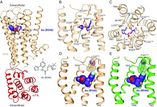
Fig. 4.
Comparison of the orthosteric binding sites of M2R and M3R. (A and B) Crystal structure of M3R in complex with the selective antagonist 6o (BS46). (A) Overall structure of the M3R/mT4L/6o (BS46) complex. (B and C) Binding-pocket residues of M3R interacting with 6o (BS46). (D and E) Interaction of 6o (BS46) with a nonconserved position in the second extracellular loop (ECL2) of M2R and M3R. (D) Crystal structure shows an interaction of the fluorine group of 6o (BS46) with Leu225 in the ECL2 of M3R. (E) Superimposed structure of M2R on the M3R/6o (BS46) structure indicates a steric clash between Phe181 of M2R and the fluorine of 6o (BS46).

