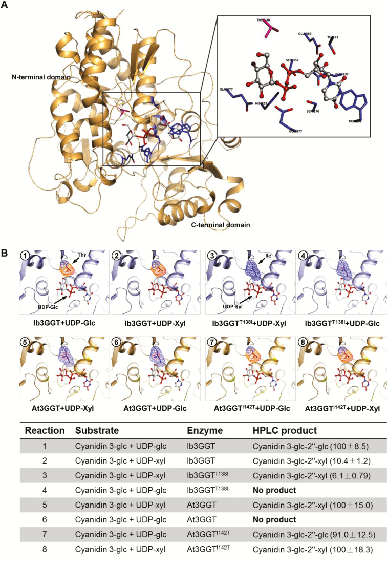
Fig. 3.
Three-dimensional modeling of Ib3GGT and At3GGT interactions with sugar donors and glycone acceptors. (A) Active center of Ib3GGT showing the key amino acid residues for the sugar donor and acceptor positions. (B) Illustrations of docking of sugar donors and a glycone acceptor in the binding pocket of wild-type and mutant Ib3GGT and At3GGT. The performance of their reactions using cyanidin 3-O-glucoside with the sugar nucleotides UDP-glucose or UDP-xylose is shown in the bottom panel. The percentage of relative enzyme activity is indicated in parentheses.

