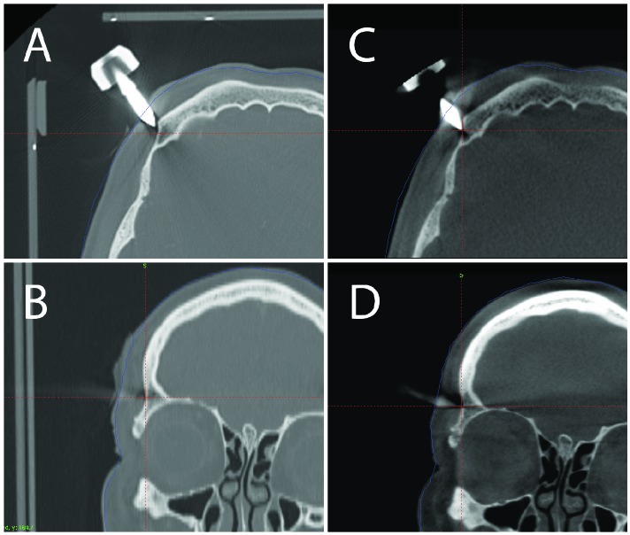
Figure 2.
Treatment planning for a left cerebellar metastasis. (A) Axial view of one of the frame pins on the stereotactic fan beam CT. (B) Coronal view of one of the frame pins on the stereotactic fan beam CT. (C) Axial view of the same pin on the pre-treatment CBCT. (D) Coronal view of the same pin on the pre-treatment CBCT. Comparison of the stereotactic fan beam CT with the pretreatment CBCT shows that the pin was not securely anchored with an accompanying frame shift.

