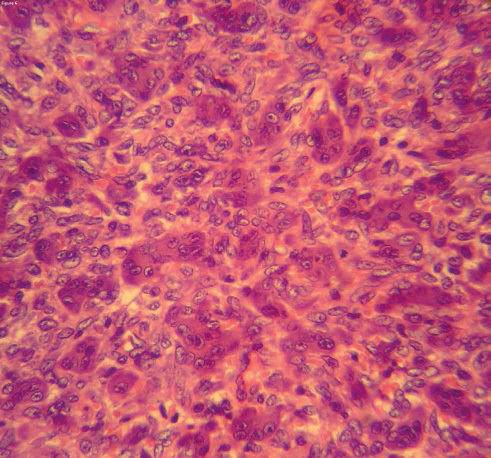
Fig. 6.

High-magnification histopathology study demonstrates two cellular populations: mononuclear cells and osteoclast-like giant cells. The giant cells were evenly distributed throughout the lesion. The mononuclear cells were oval shaped with nuclei similar to those in giant cells. A few mitotic activities are present in mononuclear cells. (Hematoxylin and Eosin, x 400.)
