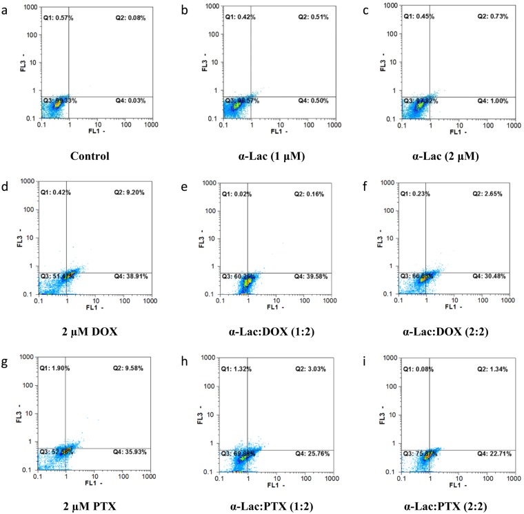
Figure 14.
Flow cytometric analysis of apoptosis induction. Effect of α-Lac, DOX, PTX, α-Lac:DOX, and α-Lac:PTX on apoptosis induction in MDA-MB-231 cells after incubation for 48 h. The cells were incubated with 1 (b) and 2 (c) μM α-Lac, 2 µM DOX (d), 1:2 ratio of α-Lac:DOX (e), 2:2 ratio of α-Lac:DOX (f), 2 μM PTX (g), 1:2 ratio of α-Lac:PTX (h), and 2:2 ratio of α-Lac:PTX (i). The presented graphs are representative of the flow cytometric output. The control cells were also presented to compare (a).

