FIGURE 5.

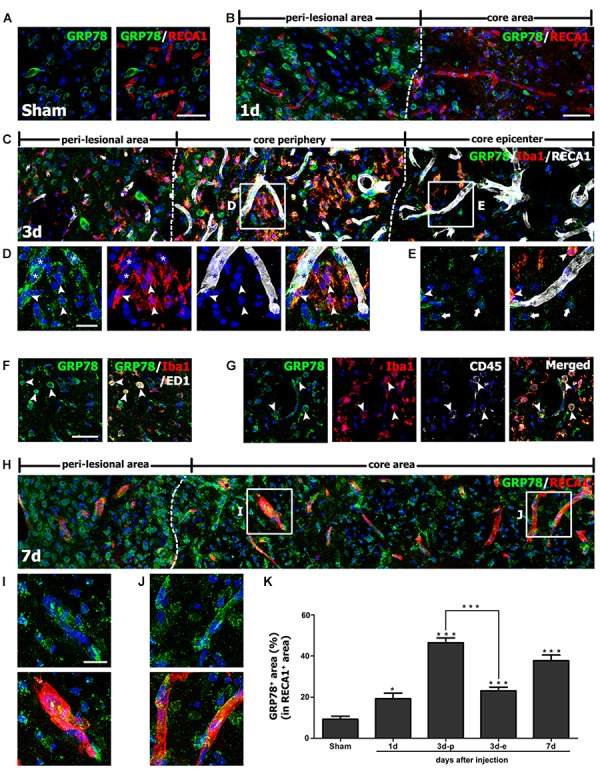
Characterization of glucose-regulated protein (GRP78) expression associated with the vasculature in control and lesioned striata after 3-nitropropionic acid injection. (A) Double-labeling of GRP78 and the endothelial cell marker RECA1 in control striatum, showing that GRP78 immunoreactivity is negligible or absent within vessels. (B) Double-labeling of GRP78 and RECA1 1 day after lesion induction, showing that GRP78 immunoreactivity within vessels is negligible or very weak in the lesion core (right side of the broken line). (C–E) Lower- (C) and higher- (D,E) magnification views of sections triple-labeled for GRP78, Iba1, and RECA1 3 days after lesion induction. The boxed areas of the lesion periphery and the epicenter in C are enlarged in D and E, respectively. Notably, the lesion periphery is heavily infiltrated by GRP78/Iba1 double-labeled microglia/macrophages (arrowheads in D), while only some Iba1-positive microglia/macrophages can be detected in the epicenter (arrowheads in E). In addition, GRP78 expression is detectable in association with most of the vasculature (asterisks in D) in the lesion periphery. Arrows in E indicate presumptive dying or dead neurons that are devoid of significant GRP78 expression. (F,G) Triple-labeling of GRP78, Iba1, and either ED1 or CD45 on day 3 after lesion induction, showing that GRP78 is expressed in nearly all of ED1- (arrowheads in F) or CD45-positive cells (arrowheads in G), corresponding to only a small fraction of the GRP78/Iba1 double-labeled cells. Notably, these triple-labeled cells are frequently associated with blood vessels. (H) Double-labeling of GRP78 and RECA1 at 7 days after lesion induction, showing that GRP78 expression is evenly distributed throughout the lesion core (right side of the broken line). (I,J) Higher magnification images of the boxed areas in H, showing that GRP78 immunoreactivity is localized to nearly all vessels in both the lesion periphery (I) and epicenter (J). (K) Quantitative temporal analysis of the proportion of vascular areas occupied by GRP78 immunoreactivity, within all RECA1-positive vessels. This proportion increases progressively by day 1, and then decreases slightly on day 7. Note that the vascular area covered by GRP78 is significantly higher in the lesion periphery than in the epicenter on day 3. The data are expressed as the mean ± standard error of the mean. ∗P < 0.05 and ∗∗∗P < 0.001 vs. saline-treated controls. Cell nuclei are stained with 4′,6-diamidino-2-phenylindole. Scale bars represent 50 μm for A–C, F–H; and 20 μm for D, E, I, J.
