Figure 5.
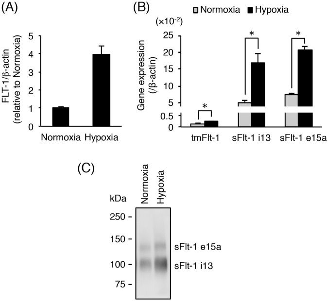
Hypoxia-induced up-regulation of sFlt-1 secretion in primary cytotrophoblasts. Thawed primary cytotrophoblasts were cultured for 16 h and then incubated for 24 h under normoxic or hypoxic conditions. Conditioned media were collected and analyzed for secretion of sFlt-1 proteins by Western blot analysis. (A) The mRNA expression of all Flt-1 transcripts (FLT-1) in cytotrophoblasts was assessed by quantitative real-time PCR analysis using β-actin mRNA as a reference. Results are represented as a fold change relative to cells under normoxic conditions. (B) The mRNA expression levels of three Flt-1 splice variants (tmFlt-1, sFlt-1 i13, and sFlt-1 e15a) in cytotrophoblasts. Results are represented as a ratio relative to the expression of β-actin mRNA. (C) Secreted sFlt-1 proteins in conditioned media were concentrated by heparin-sepharose beads and then subjected to Western blot analysis with anti-human Flt-1 N-terminal antibody. Uncropped image of Western blot is presented in Supplementary Fig. S5. All values are represented as the means ± SD (n = 3). Asterisks indicate a significant difference (P < 0.05).

