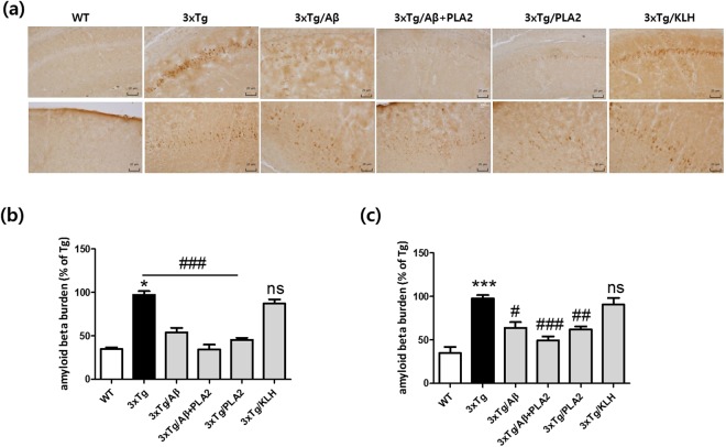
Figure 5.
Immunohistochemical staining depicting the effects of bvPLA2 treatment on Aβ burdens in the brains of 3xTg-AD mice. Aβ burdens were determined by immunohistochemical staining with anti-Aβ antibodies. (a) Representative images of Aβ staining in the hippocampal CA1 (top) and cortex (bottom) regions. Scale bars 20 µm. Bar graphs of data pertaining to the effects of bvPLA2 treatment on Aβ deposition in (b) hippocampal CA1 and (c) cortex regions. The data are shown as the mean ± SEM. ***P < 0.001 vs. WT; #P < 0.05, ##P < 0.01, ###P < 0.001 vs. 3xTg.

