FIGURE 3.
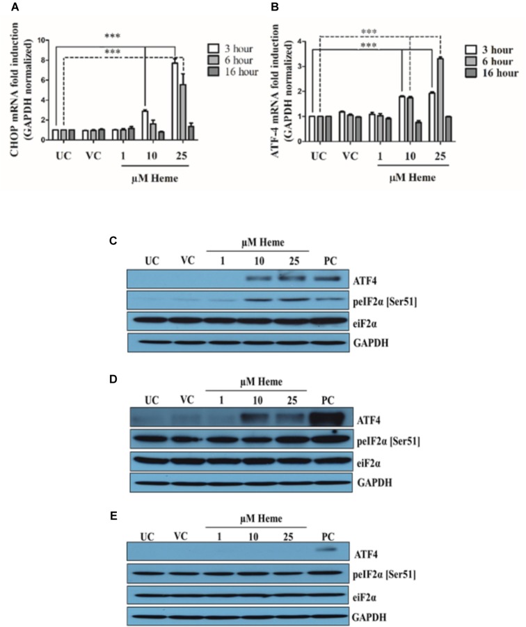
Heme activates the PERK/ATF4/CHOP arm of ER stress in a time- and dose-dependent manner. HAoSMCs were treated with various doses of heme (1,10, and 25 μM) or corresponding vehicle solution to highest heme dose (25 μM) in serum-free DMEM for 60 min, then medium was changed to DMEM with 10% FCS and antibiotics. ER stress markers were measured after 3, 6, or 16 h. Thapsigargin (1 μM) treated cells were used as positive control (A–E). (A,B) Relative expressions of CHOP (A) and ATF4 (B) mRNA levels were determined by qRT-PCR, normalized to GAPDH and compared to the untreated controls at each time points. UC, untreated control; VC, vehicle control; PC, positive control, thapsigargin treated. Results are presented as mean ± SD of five independent experiments performed in duplicates. ∗∗∗p < 0.001. (C–E) Representative Western blots of whole cell lysates from five independent experiments are shown representing eIF-2 phosphorylation and ATF4 protein levels (C) 3, (D) 6, and (E) 16 h after the heme treatment. UC, untreated cells; VC, vehicle control cells; PC, positive control cells.

