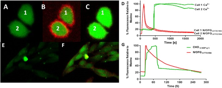
Figure 4.

Live cell binding of N/OFQATTO594 and subsequent stimulation in CHOhNOPGαqi5 cells. Fluo4‐AM loaded CHOhNOPGαqi5 cells at 4°C (A) are incubated with 100 nM N/OFQATTO594 after 30 s (B) leading to release of calcium (cells labelled 1 and 2) (B and C). The process is demonstrated in the Supporting Information Video S1. This video has been created through ImageJ (eight frames per second), with N/OFQATTO594 added after 15 s, with the entirety of the video covering approximately 4.5 min. (D) A representative figure demonstrating N/OFQATTO594 binding to CHOhNOPGαqi5 cells at 4°C and the increase in Ca2+. Note: red is N/OFQATTO594 binding and green is Ca2+. Fluo4‐AM loaded CHOhNOPGαqi5 cells at 37°C (E) are incubated with 100 nM N/OFQATTO594 after 30 s, which leads to a prompt increase in binding and calcium (small field of cells; F). (G) A representative figure demonstrating binding of N/OFQATTO594 at 37°C and increase in Ca2+. Note: red is N/OFQATTO594 binding and green is Ca2+. These data are representative of n = 5.
