Figure 6.

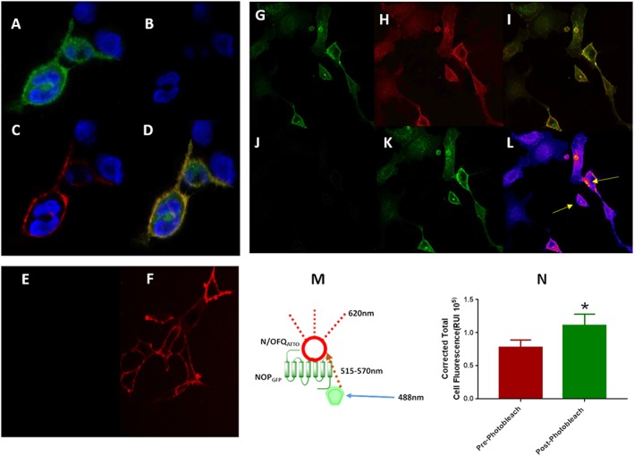
N/OFQATTO594 can be activated when in close proximity to GFP attached to the NOP receptor (FRET), as described in the cartoon in the middle of the figure (M). (A) Visualizes the NOP‐GFP receptors stimulated by the 488 nm laser in the green filter window. (B) Demonstrates no ‘leak’ of fluorescence into the red window when using the green laser in the absence of N/OFQATTO594. Upon addition of 100 nM N/OFQATTO594, the ligand is stimulated through FRET pairing (C) to fluoresce when in close proximity to the NOPGFP receptors. Signals are overlapped in (D). (E) In order to confirm FRET pairing, the 488 nm laser was used in conjunction with HEKhNOP cells (no fluorescent linker present) to again demonstrate lack of activation of N/OFQATTO594. (F) Binding of N/OFQATTO594 was confirmed using 594 nm laser. Photobleaching of the acceptor molecule is a further method to confirm FRET pairing. HEKhNOPGFP cells (G) were labelled with 100 nM N/OFQATTO594 (H) with binding of the receptor‐ligand complex shown as a composite image (I). N/OFQATTO was exposed to 594 nm light until photobleaching was achieved (J) at which point changes in NOPGFP fluorescence were measured (K) with the heatmap indicating increases of fluorescence shown and highlighted by arrows (blue: low and red: high) (L). (N) Average increase in NOPGFP fluorescence after photobleaching of N/OFQATTO594; P < 0.05 Students t‐test. Data are the mean of eight experiments.
