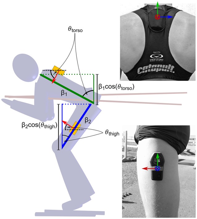
Figure 1.

Positioning of the two IMUs on the athletes' body and motivation for the frontal area model described in Equation 5. One IMU was positioned approximately at the level of the third thoracic vertebra, the other was taped laterally on the thigh approximately 10 cm inferior to the trochanter. The axes were aligned with the x-axis (blue) in the mediolateral direction and the y-axis (red) in the anterior direction. The z-axis (green) was aligned with gravity when the athletes were in a standing posture, as described in the text. The frontal areas of the torso and thighs scale approximately with the cosine of the pitch angles θ k = atan(ay, k/az, k), where ay, k and az, k are the smoothed accelerometer outputs along the y and z-axis respectively. k represents the thigh or torso location.
