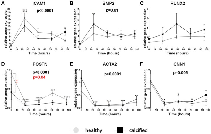
Figure 2.
Relative gene expression of intracellular adhesion molecule 1 (ICAM1) (A), bone morphogenetic protein 2 (BMP2) (B), runt-related transcription factor 2 (RUNX2) (C), periostin (POSTN) (D), α-smooth muscle actin (ACTA2) (E), calponin 1 (CNN1) (F) in valve interstitial cells isolated from healthy (n = 4, gray) and calcified (n = 4, black) valves cultured on collagen I coating, stimulated with LPS and collected on different time points (24, 48, 72, and 96 h). Changes in gene expression in cells with LPS stimulation in relation to control cells without LPS stimulation are shown in gray (for healthy donors) and black (for calcified donors) stars. Differences in gene expression between cells from healthy and calcified donors are shown in red stars. * indicates 0.01 < p ≤ 0.05, ** indicates 0.001 < p ≤ 0.01, *** indicates 0.0001 < p ≤ 0.001, **** indicates p ≤ 0.0001. Overall p-values from two-way ANOVA indicating differences in gene expression between cells from healthy and calcified donors are shown in red. Overall p-values from two-way ANOVA indicating effect of the time on gene expression are shown in black. Data presented as mean ± SEM. p-values < 0.05 were considered statistically significant.

