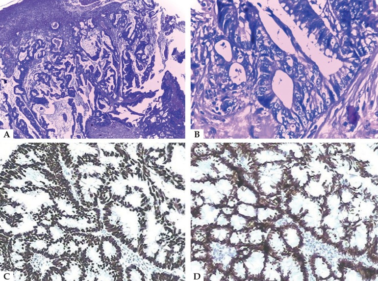
Figure 2.
Histopathological exam. A - Metastatic adenocarcinoma - neoplasm composed of well-formed ductal structures that had infiltrated the dermis, whose epithelium was composed of columnar cells of pleomorphic vesicular nuclei, with more than one nucleolus and frequent atypical mitotic figures. The fibrovascular stroma showed erythrocyte extravasation and a mixed inflammatory reaction of lymphocytes, neutrophils and eosinophils, fibrosis and ectatic vessels (Hematoxylin & eosin, x100);

