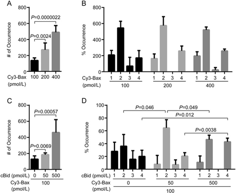
Figure 1.
Translocation and oligomerization of Bax in lipid bilayers with or without cBid. (A) Numbers of fluorescent spots (# of Occurrence) in the images of 100, 200 and 400 pmol/L Cy3-labelled Bax (C126S) in DPPC lipid bilayers with cardiolipin (93% DPPC and 7% cardiolipin). Error bars indicate the standard errors, n=6. Data analyzed by an unpaired one-tailed t test. (B) Percentage of occurrence (% Occurrence) of different oligomeric species in the images (A). Error bars indicate the standard errors, n=3. (C) Numbers of fluorescent spots (# of Occurrence) in the images of 100 pmol/L Cy3-labelled Bax (C126S) with 50 and 500 pmol/L cBid in DPPC lipid bilayers with cardiolipin (93% DPPC and 7% cardiolipin). Error bars indicate the standard errors, n=6. Data analyzed by an unpaired one-tailed t test. (D) Percentage of occurrence (% Occurrence) of different oligomeric species in the images (C). Error bars indicate the standard errors, n=3. Data analyzed by an unpaired one-tailed t test.

