Figure 3.
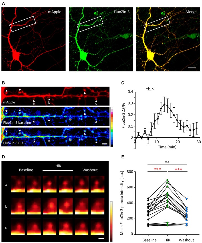
Depolarization induces a transient and reversible increase of zinc in postsynaptic spines. (A) A young hippocampal neuron (DIV 14) transfected with mApple (left, red) and loaded with FluoZin-3 (middle, green). mApple was used as a morphological marker to indicate dendrites and postsynaptic spines. Yellow indicates clear colocalization in the merge image (far right). White boxes mark the dendrite shown in (B). Scale bar: 10 μm. (B) Straightened dendrite with mApple (top) and FluoZin-3 at baseline (middle) and during depolarization conditions [bottom; 90 mM KCl (HiK), 120–180 s]. White arrowheads mark spines being quantified in (C) and shown in (D; indicated by a–c). Scale bar: 5 μm. (C) Time course of changes in FluoZin-3 (ΔF/Fo) with depolarization for spines in (B). Black line indicates when depolarization was applied (HiK’). Mean ± SEM for N = 6 spines in one dendrite, shown in (B). (D) FluoZin-3 fluorescence changes in individual spines (a–c) under baseline, HiK (white line) and washout conditions indicated in (B). Scale bar: 1 μm. (E) Effects of depolarization on FluoZin-3 in spines of individual hippocampal neurons averaged across baseline, during HiK and washout conditions (one-way ANOVA followed by Sidak’s multiple comparisons, N = 17 dendrites from five neurons from four culture preps, n.s. p > 0.05, ***p < 0.001).

