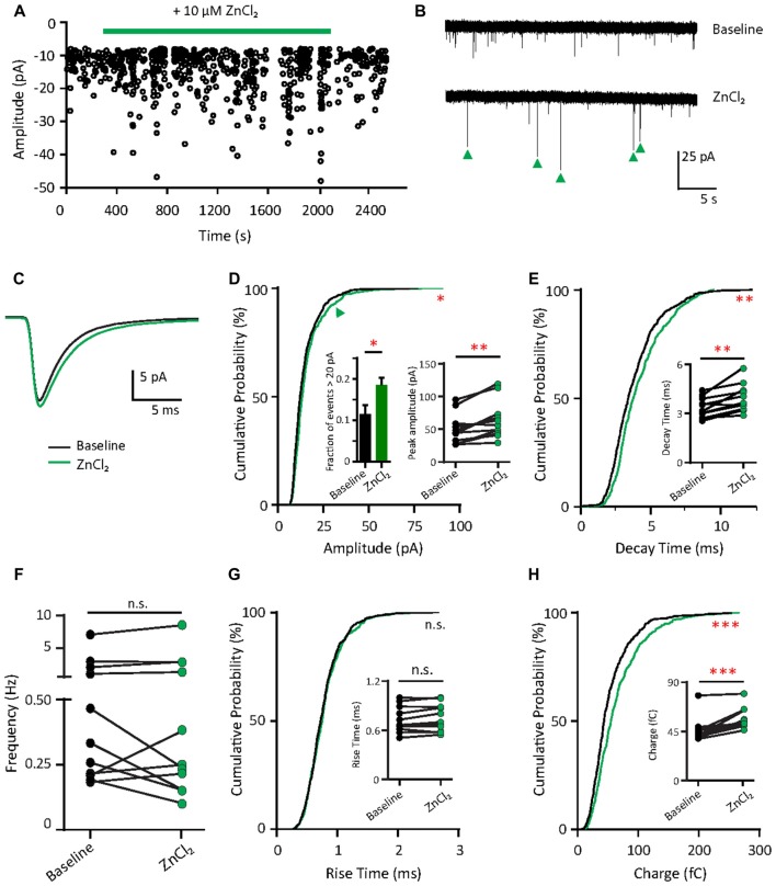
Figure 4.
Zinc treatment enhances synaptic efficacy of AMPAR mEPSCs in young neurons. (A) AMPAR mEPSCs were recorded from a young hippocampal neuron (DIV 11). Green bar: 10 μM ZnCl2 application. (B) AMPAR mEPSC recording traces from the neuron shown in (A) at baseline (top) and during ZnCl2 (bottom). Green arrowheads mark the events with larger amplitude during ZnCl2 treatment. (C) Ensemble-averaged mEPSCs from the baseline and zinc conditions measured in the same neurons (baseline: black, ZnCl2: green, N = 11 cells from seven culture preps, which applies to all subsequent panels unless otherwise specified). (D,E,G,H) Cumulative probability histograms of amplitude (D), decay time (E), rise time (G) and charge (H) of isolated events from baseline (black) and ZnCl2 (green) conditions (K.S. test, N = 600 events. n.s. p ≥ 0.005, *p < 0.005, **p < 0.001, ***p < 0.0001). First inset in (D): summary graph showing the difference of fraction of events with amplitude >20 pA between baseline and ZnCl2 conditions. All other insets show per-cell-basis pairwise comparison of amplitude (D), decay time (E), rise time (G) and charge (H) between baseline and ZnCl2 conditions (Wilcoxon test, n.s. p ≥ 0.05, **p < 0.01, ***p < 0.001). (F) Pairwise comparison of AMPAR mEPSC frequency between baseline and ZnCl2 conditions (Wilcoxon test, n.s. p ≥ 0.05).

