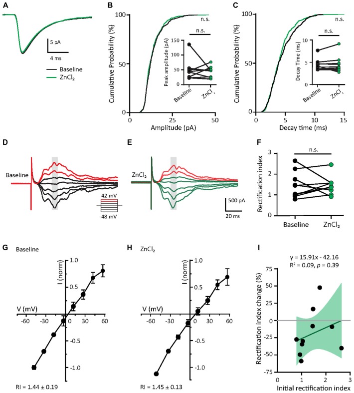
Figure 6.
Zinc treatment does not affect miniature or evoked AMPAR EPSCs in mature neurons. (A) Ensemble-averaged mEPSCs from the baseline and zinc conditions recorded in mature neurons (baseline: black, ZnCl2: green, N = 10 cells). (B,C) Cumulative probability histograms of amplitude (B) or decay time (C) of isolated events from baseline and ZnCl2 conditions (K.S. test, N = 400 events from 10 cells from four culture preps per condition, n.s. p ≥ 0.05). Insets display per-cell-basis pairwise comparison of amplitude (B) and decay time (C; Wilcoxon test, N = 10 cells, n.s. p ≥ 0.05). (D,E) AMPAR-mediated EPSCs (averaged from three trials) evoked at various holding potentials [diagram (middle)] from a mature hippocampal neuron (DIV 18) during baseline (D) and after 10 min of 10 μM ZnCl2 application (E). Red traces correspond to the current response at the two most positive holding potentials. Gray shaded bars mark the regions used for calculating peak current amplitude in (G,H). (F) Summary graph showing effect of ZnCl2 application on RI of mature neurons (Wilcoxon test, N = 10 cells from three culture preps, which applies to all subsequent panels unless otherwise specified; n.s. p ≥ 0.05). (G,H) Normalized current-voltage relationship (mean ± SEM) of the pooled data recorded from the same neurons at baseline (G) and ZnCl2 treatment (H). (I) Relationship between the initial RI (during baseline) and the magnitude of RI change with ZnCl2 application (Pearson correlation, n.s. p ≥ 0.05). Green shaded area indicates the 95% confidence interval.

