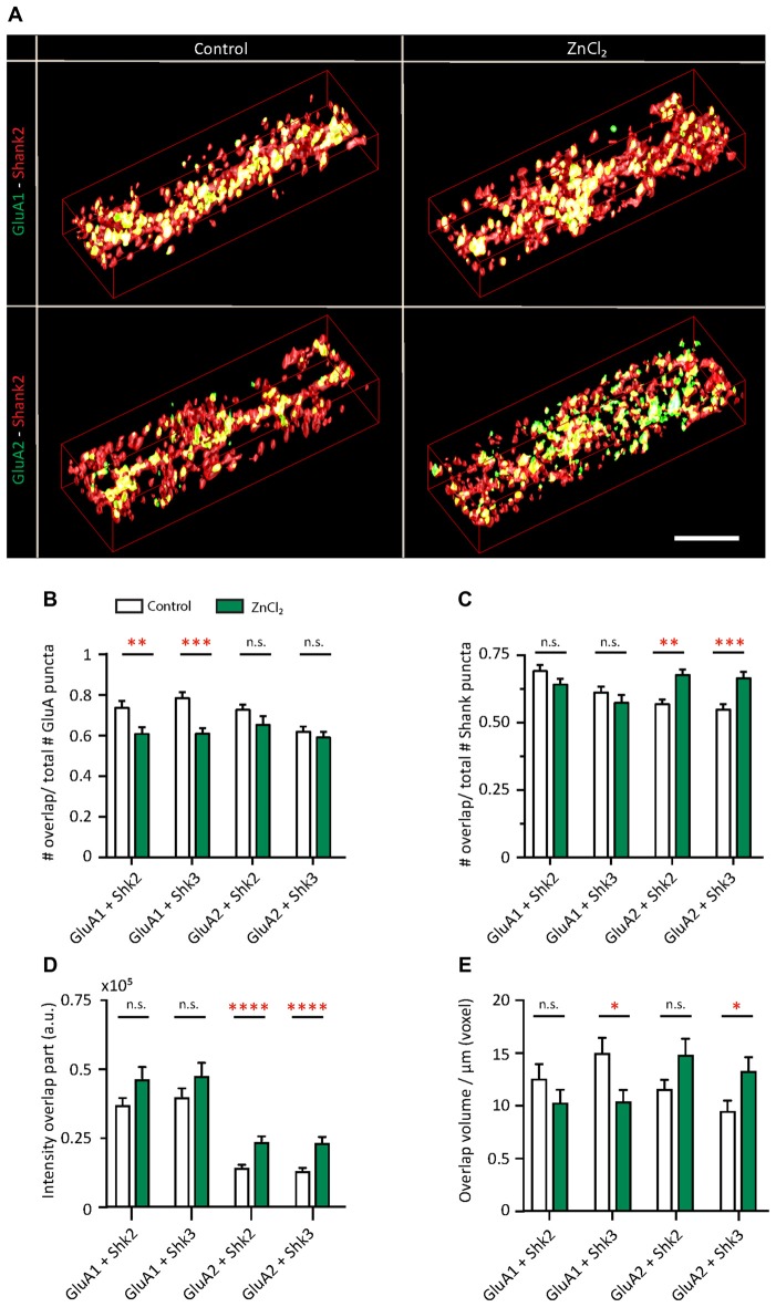
Figure 8.
Zinc treatment alters the colocalization between AMPAR subunits and Shank. (A) Straightened dendrites from young hippocampal neurons (DIV 14) treated with control (left) or 10 μM ZnCl2 (right). Neurons were live surface labeled for GluA1 (green; top) or GluA2 (green; bottom) before being fixed and co-immunostained for both Shank2 (red) and Shank3 (not shown). Yellow indicates colocalization. Scale bar: 4 μm. (B–E) Three-dimensional colocalization analysis of pairwise puncta overlap using IMFLAN3D as measured by the fraction of overlap (B,C), intensity (D) and volume (E) for baseline and ZnCl2 conditions (mean ± SEM) [Mann-Whitney test, for GluA1 + Shank3 or Shank2, N = 19 (control) and 22 (ZnCl2) dendrites from 10 to 14 neurons from two culture preps; for GluA2 + Shank3 or Shank2, N = 24 dendrites from 10 to 14 neurons from two culture preps; for Shank2 + Shank3, N = 43 (control) and 46 (ZnCl2) dendrites from 20 to 25 neurons from four culture preps; n.s. p ≥ 0.05, *p < 0.05, **p < 0.01, ***p < 0.001, ****p < 0.0001].

