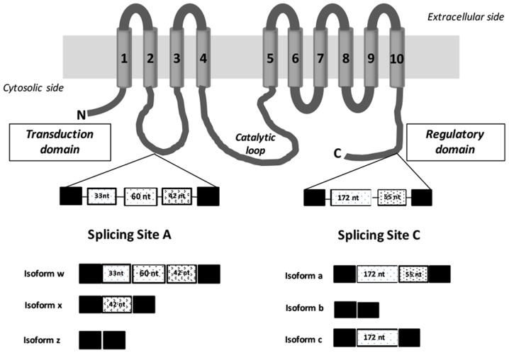
Figure 1.
Diagram illustrating the structure of human plasma membrane calcium ATPase 2 and its splice isoforms. The protein consists of 10 transmembrane domains numbered and indicated in gray tubes. The two alternative splicing sites are depicted in the figure, the splice sites A (N-terminal domain) and C (C-terminal tail) within the regulatory domain (calmodulin-binding domain). The intracellular loop connecting TM4 and TM5 is termed the catalytic loop, and contains the phosphorylation and ATP-binding sites. Alternative splicing of human PMCA2 pump at site A located between transmembrane domains 2 and 3 generates three isoforms w, z and x. At site C three isoforms a, b or c could be generated by alternative splicing in the C-terminal region. Constitutively spliced exons are depicted as black boxes and alternatively inserted exons are depicted in white with different patterns. nt, number of nucleotides; N, N-terminal domain; C, C-terminal domain.

