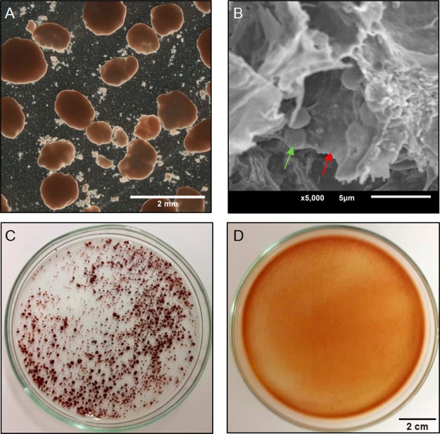
Figure 1.
A) Optical microscope image of anammox granular sludge from the WWTP. B) Scanning electron microscope image of the inside of a broken granule where bacteria (green arrow) can be seen, embedded in the EPS matrix (red arrow). C) Anammox granules before and D) after incubation for 5 h in 0.1 M NaOH.

