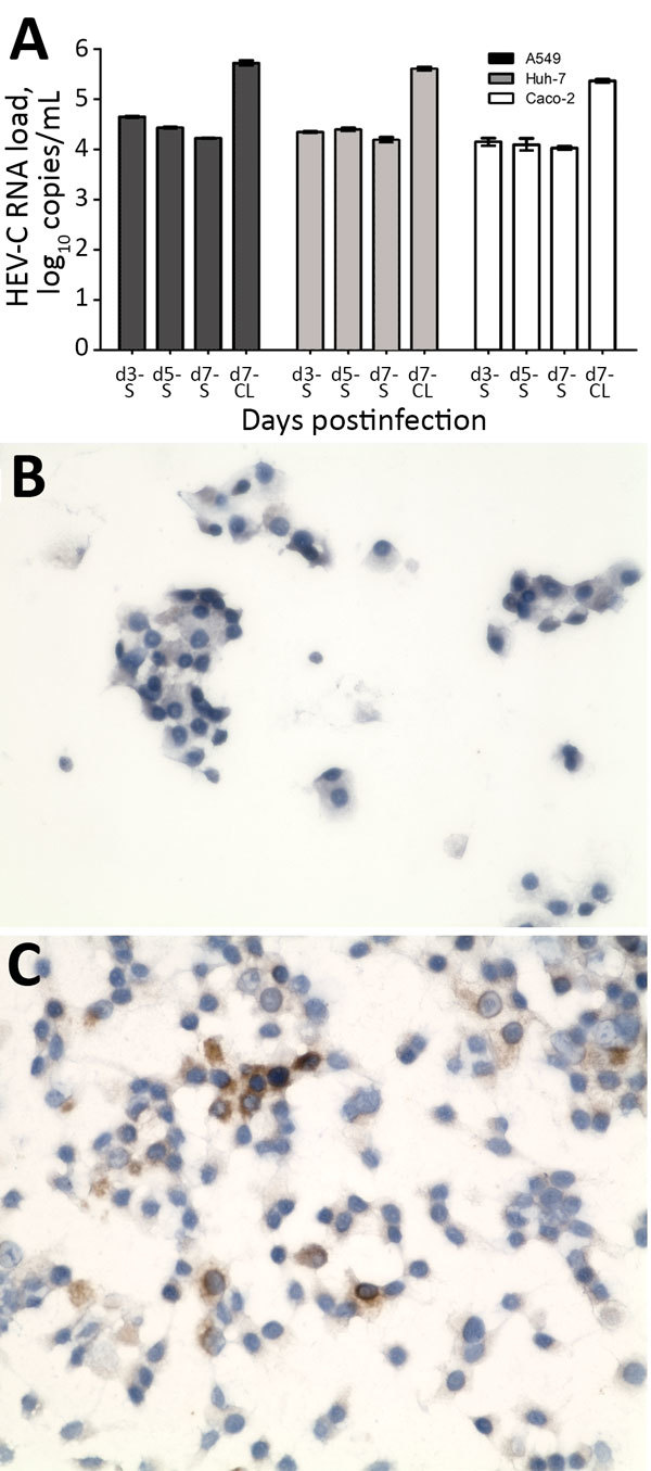
Figure 5.

Isolation of HEV-C from 56-year-old male patient’s feces in cell culture, Queen Mary Hospital, Hong Kong. A) HEV-C RNA loads in culture S and day-7 CL of A549, Huh-7, and Caco-2 cell lines after inoculation by patient’s filtered fecal suspension. Mean of 3 replicates; error bars indicate SEM. B) Uninfected A549 cell monolayer stained with anti–HEV-C polyclonal antiserum. C) Infected A549 cell monolayer stained with anti–HEV-C polyclonal antiserum. Original magnification ×400. CL, cell lysate; HEV, hepatitis E virus; S, supernatant.
