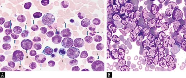
Figure 1. A) Showing hyposegmented basophils (1, 2, 6), binuclear erythroblast (3), giant forms of basophilic bands (4, 5), large eosinophilic myelocyte (7), erythroblast with dysplastic nucleus (8), giant basophilic hypogranular metamyelocyte (9), giant binuclear basophilic metamyelocyte (10), basophilic myelocyte (11), neutrophilic band (12), segmented neutrophil (13) in chronic phase of primary chronic basophilic leukemia (Wright’s stain, 100x); B) Demonstrating red color (metachromatic) granular cytoplasmic staining in 70% nucleated cells of the bone marrow (toluidine blue stain, 100x).