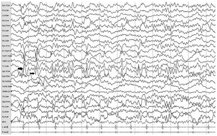
Figure 2.
Typical electroencephalogram of a patient with hippocampal sclerosis. Traces exhibit multiple continuous slowing epileptic discharges (indicated with arrows) from the left anterior temporal electrodes (Sp1 and T1) during the interictal period, which indicate a potential onset zone located in the mesial or lateral anterior temporal lobe.

