
Figure 5.
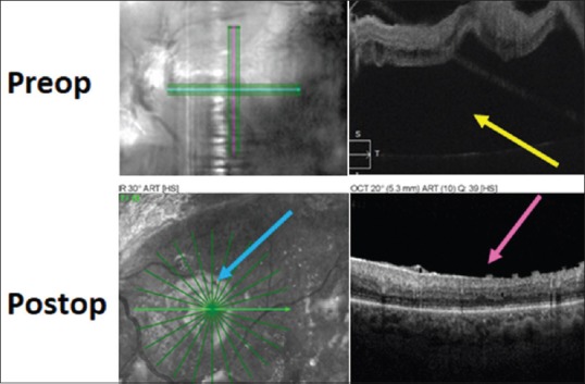
Pre-operative (top) and post-operative (bottom) optical coherence tomography images in the case of traction retinal detachment involving the macula shown in Figures 1 and 2. The yellow arrow shows subfoveal fluid pre-operatively and the fluid is gone post-operatively. The pink arrow shows loss of the foveal depression in the post-operative macula, which is a common finding. The blue arrow shows the light reflex at the silicone oil-retina interface