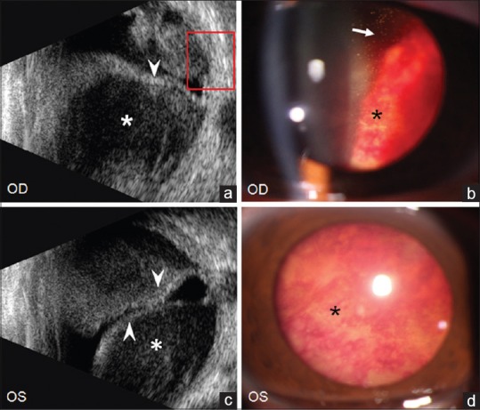
Figure 1.

The initial ultrasound (a and c) and slit-lamp photography (b and d) of bilateral eyes. Many gray and white particles (arrows in b) in vitreous body, diffuse retinal hemorrhages (black asterisk in b and d), complete retinal detachments (arrow heads in a and c) and extensive subretinal hemorrhage (white asterisk in a and c) were observed in both the eyes
