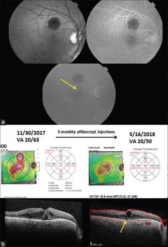
Figure 3.

Images of a 77-year-old man with type 2 diabetes mellitus, who developed diabetic macular edema of the left eye that reduced visual acuity to 20/63. (a) Red free fundus photography shows microaneurysms and one large blot hemorrhage above the fovea. Fluorescein angiography shows multiple hyperfluorescent microaneurysms in the mid-phase, and late leakage above the fovea in the late frame. (b) The spectral domain-optical coherence tomography on November 30, 2017 shows center-involved diabetic macular edema and subfoveal fluid. After 5 monthly aflibercept injections, the edema has decreased, but persistent edema is present. An ellipsoid zone defect (yellow arrow) is apparent
