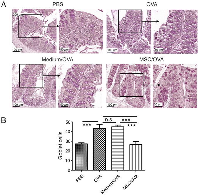
Figure 6.
Goblet cell staining in the colon. Mice were divided into PBS, OVA, Medium/OVA and MSC/OVA treatment groups. (A) Representative images of the goblet cells in the colon, stained using periodic acid-Schiff stain. Scale bar, 100 and 50 µm. (B) Quantification of the goblet cells in the fields under the microscope. hUC-MSC, human umbilical cord-derived mesenchymal stem cell; OVA, ovalbumin; Medium/OVA, group challenged with OVA and administered Dulbecco's modified Eagle's medium/nutrient mixture F12; MSC/OVAOVA/MSC, group challenged with OVA, intraperitoneal injection of hUC-MSCs and oral gavage of MSC culture supernatant. ***P<0.001. ns, not significant.

