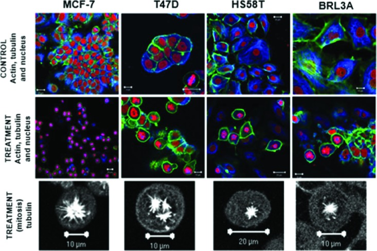
Figure 4.
Formation of atypical mitotic spindles in 2,3,9-trimethoxypterocarpan-treated cells. Actin (green), tubulin (blue) and nuclei (red) are labeled in breast carcinoma and nontumor (BRL3A) cells treated or not treated with the compound. Note the normal arrangement of tubulin and actin fibers in the control cells; the formation of monopolar spindles in MCF-7, HS58T and BRL3A cells; and the formation of tripolar spindles in T47D cells. Figure from Militão et al. 97.

