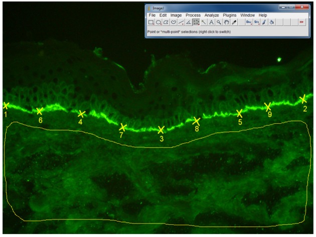
Figure 7.

Algorithm of measurement of fluorescence intensity in direct IF microscopy images, in patients with BP (n = 27 patients). The fluorescence intensity was assessed by two independent blinded observers using Image J software, 1.47v version. The fluorescence intensity at the basement membrane was analyzed by selecting 50 points (using Multi-point tool), each point being situated at the half distance between two previous points. Background fluorescence was evaluated by selecting the whole dermis area, using “Freehand selections” tool. In order to obtain the mean value and standard deviation of the fluorescence intensity for the 50 points on the basement membrane and dermis background we have used the Analyze-Measure-Summarize tool.
