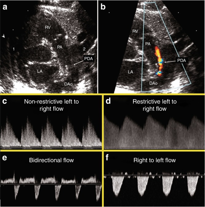
Fig. 2.

PDA 2D, color Doppler image and Doppler flow patterns. The top panels demonstrate the PDA in 2D (a) and color Doppler (b). c Pulsatile or non-restrictive pattern: characterized by a left to right (LtR) shunt with an arterial waveform and high peak systolic velocity: end-diastolic velocity ratio. d Restrictive pattern: characterized by high systolic and diastolic velocity, and low peak systolic velocity: end-diastolic velocity ratio. e Bidirectional pattern: elevated pulmonary pressures equal to or near systemic. f Right to left (RtL) flow: supra-systemic pulmonary pressures
