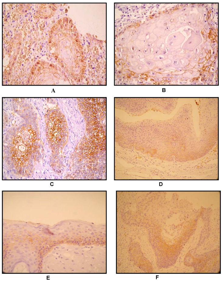
Figure 2.
A) Keratinizing squamous carcinoma. GLUT1, prostromal staining pattern. Note positivity at periphery of tumour nest, in non-keratinizing basaloid cells, and loss of GLUT1 expression accompanying keratinization at the centre of tumour nest, 200×; B) 400×; C) Non - keratinizing poorly differentiated carcinoma, 100×. In the absence of squamous differentiation/keratinization: GLUT1 displays an antistromal pattern, suggestive of hipoxia-driven GLUT1 induction. D) and E) Basal staning pattern with the superficial layers showing little or no GLUT1 staining in the normal epithelium; F) GLUT1 immunostain (arrow). Squamous intraepithelial neoplasia, non- staining of basal layer.

