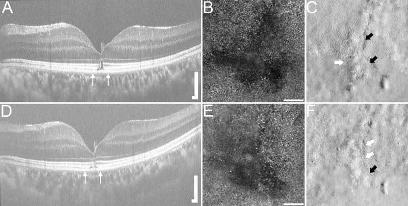
Figure 2.
Multimodal imaging of the left eye for subject WW_0923 at 22 months (A,B,C) and 52 months (D,E,F) after trauma. Macular horizontal SD-OCT line scan revealed a persistent, focal outer lamellar defect with EZ and IZ disruption (A), which improved over time (D). Thin white arrows in (A) and (D) indicate area of AOSLO imaging. Confocal macular AOSLO imaging demonstrated a large anchor-shaped area of non-waveguiding photoreceptors (B), which is smaller in size on follow-up (E). Split-detector AOSLO images of the same location revealed a similar anchor-shaped disruption with both regions of intact inner-segment photoreceptors (white arrow) and areas devoid of inner-segment structure (black arrows) (C). Split-detector AOSLO imaging at 52 months post-trauma (F) showed inner-segment photoreceptor structure in areas previously without structure (F, white arrows), although focal areas of absent inner-segment structure still exist (F, black arrow). A and D: scale bar=200 µm; B, C, E and F: scale bar=100 µm. AOSLO, adaptive optics scanning light ophthalmoscopy; EZ, ellipsoid zone; IZ, interdigitation zone; SD-OCT, spectral domain optical coherence tomography.

