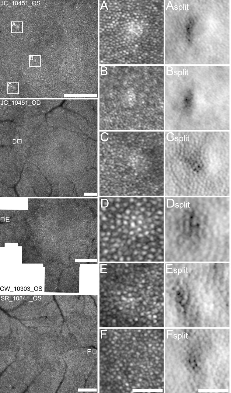
Figure 4.
Confocal and split-detector AOSLO subclinical photoreceptor mosaic anomalies. Focal regions of abnormal waveguiding photoreceptors were found in confocal AOSLO foveal mosaics in subjects JC_10451 OD & OS, CW_10303 OS and SR_10341 OS (white boxes). Magnified views of these regions show focal hyper-reflective waveguiding cones with confocal AOSLO imaging (A–F) and focal mounding of photoreceptor inner-segment structure on split-detector AOSLO imaging (Asplit–Fsplit), ranging from 25 µm to 40 µm in size. Montages: scale bar=300 µm. A–Fsplit: scale bar=40 µm. AOSLO, adaptive optics scanning light ophthalmoscopy.

