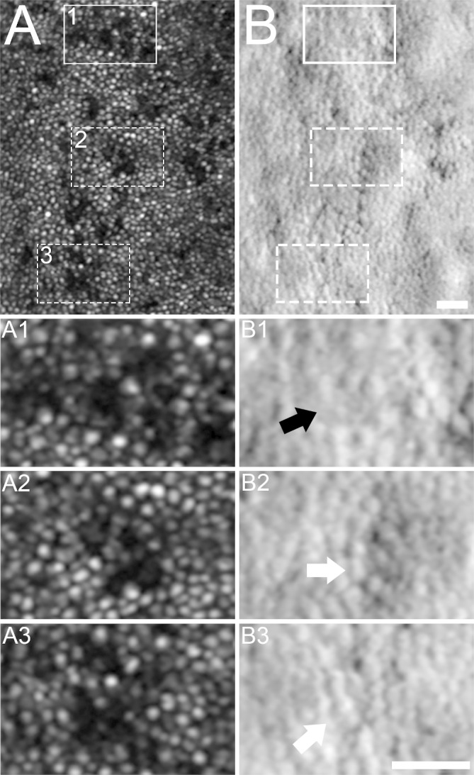
Figure 5.

Confocal and split-detector AOSLO imaging in areas of photoreceptor mosaic disruption in subject KS_0552. Foveal confocal AOSLO revealed multiple areas of non-waveguiding cone photoreceptors (A). Three of these regions (A1–A3) were compared with aligned corresponding split-detector images (B1–B3). Areas of both absent inner-segment structure (B1, black arrow) and remnant inner-segment photoreceptor structure (B2, B3, white arrows) were present in these areas of non-waveguiding confocal images. A–B3: scale bar=25 µm. AOSLO, adaptive optics scanning light ophthalmoscopy.
