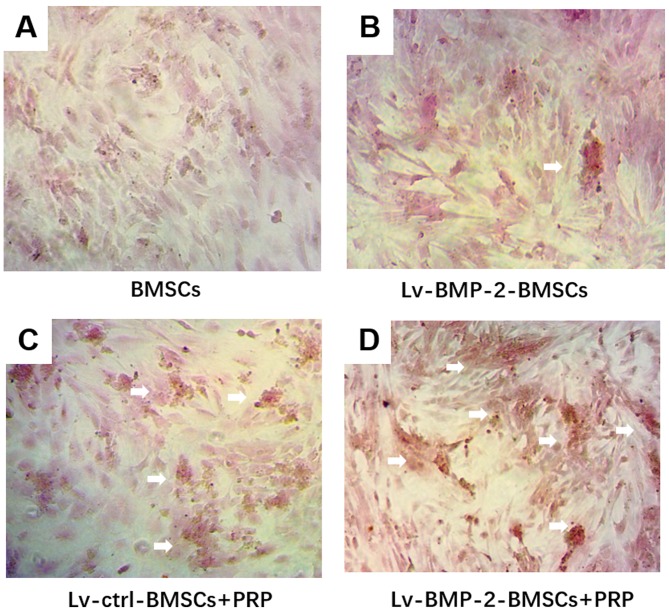
Figure 5.
Osteogenic differentiation in BMSCs. Cells were cultured with osteogenesis induction medium for 14 days followed by Alizarin red staining. Mineralized nodules in the (A) BMSCs, (B) Lv-BMP-2 BMSCs, (C) Lv-ctrl BMSCs+PRP, and (D) Lv-BMP-2 BMSCs+PRP were observed under a microscope (magnification, ×400). White arrows show mineralized calcium deposits. BMP-2, bone morphogenetic protein-2; BMSCs, bone marrow-derived stromal cells; PRP, platelet-rich plasma; ctrl, control.

