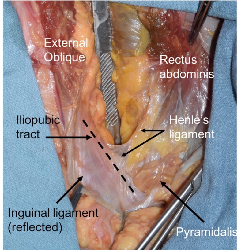
Figure 1. Right inguinal region dissected in a fresh frozen adult cadaver.
The scalpel handle is pushing down the contents of the peritoneal cavity and the inguinal ligament is cut and reflected inferiorly. The iliopubic tract is seen at the dotted line. An extension of the lateral aspect of the rectus abdominis muscle is seen as Henle’s ligament.

