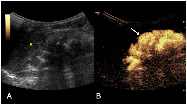
Figure 7.
(A) Two-dimensional ultrasound image and (B) contrast-enhanced voiding urosonography image of a Grade V vesicoureteral reflux on the left side. The contrast agent appeared in the pelvis and ureter with clear expansion of the pelvis and calices, and disappearance of carunclulae papillaris (arrow).

