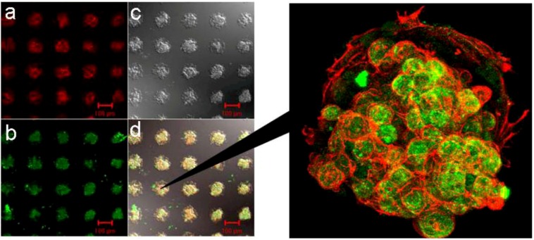
Figure 8.
Confocal laser scanning microscopy of patterned three dimensional spheroids after the double staining of F-actin and albumin, co-cultured for three weeks at 37 °C (left). Spheroids were fixed and double stained with: (a) rhodamine-conjugated phalloidin for F-actin and (b) anti-rat albumin antibody and FITC-conjugated second antibody for albumin synthesis activity. (c) Interference reflection microscopy. (d) Superimposition of (a), (b), and (c). The four images were obtained from the same view field. Scale bars are 100 μm(right) 3-D view of spheroids, underlaid with endothelial cells as a feeder layer [28].

