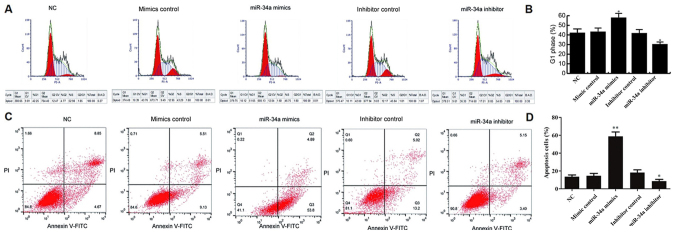
Figure 4.
miR-34a induces MCF-7 cell apoptosis and G1 phase arrest. At 48 h after cell transfection, the cell cycle distribution and apoptosis of MCF-7 cells were determined by flow cytometry. (A) Cell cycle distribution and (B) the percentage of cells in G1 phase. (C) Apoptosis of MCF-7 cells in different groups. Q2 and Q3 indicate the early and late apoptosis, respectively. (D) The percentage of apoptotic cells. *P<0.05 and **P<0.01 vs. NC group. miR, microRNA; NC, negative control.

