Figure 5.
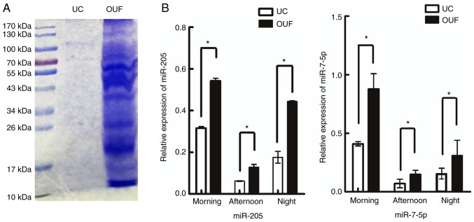
Stability and structure integrity of exosome. (A) SDS-PAGE protein pattern. Colloidal Coomassie-stained gel of exosome fractions isolated by UC and OUF. In total, 20-µl urinary exosomes were loaded per lane in the order of morning, middle and night. Sizes of reference bands are indicated on the left in kilodaltons (kDa). (B) RT-qPCR detection of miR-205 and miR-7-5p levels in urinary exosomes from health donor samples that were collected at three different time and respectively isolated by UC and OUF. Results are expressed as the mean ± standard deviation (n=3; 3 biological replicates, with 3 technical replicates each). *P<0.05. UC, ultracentrifugation; OUF, optimized ultrafiltration.

