Figure 5.
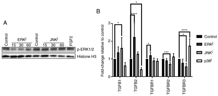
TGF-β-associated gene expression profiles induced by treatment with mitogen-activated protein kinase inhibitors. (A) CRL-2097 human dermal fibroblasts were cultured for 15-60 min in the presence or absence of 10 µM U0126 (ERKi) or 10 µM SP600125 (JNKi) and subjected to western blot analysis for p-ERK1/2. Histone H3 was used as a loading control. Fibroblasts incubated with recombinant human FGF2 for 30 min were used as a positive control for p-ERK1/2 expression. (B) CRL-2097 human dermal fibroblasts were cultured in the presence or absence of 10 µM U0126 (ERKi), 10 µM SP600125 (JNKi) or 10 µM SB202190 (p38i) until day 4. Expression levels of TGF-β-associated transcripts TGFB1, TGFB2, TGFBR1, TGFBR2 and TGFBR3 were determined relative to fibroblasts cultured under control conditions by reverse transcription-quantitative polymerase chain reaction. GAPDH expression was used as an internal control. Expression levels per transcript were compared using an one-way analysis of variance and post-hoc Holm-Sidak analysis. *P<0.05, ***P<0.001 and ****P<0.0001. There were 6 biological replicates/condition. Error bars represent the standard deviation. TGFB1, transforming growth factor-β1; TGFBR1, TGF-β receptor 1; p-ERK, phosphoextracellular signal-regulated kinase; JNK, c-Jun N-terminal kinase; FGF2, fibroblast growth factor 2; i, inhibitor.

