Figure 6.
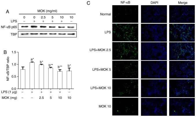
Effects of MOK extract on the expression of NF-κB induced by LPS in RAW 264.7 cells. Graded concentrations of MOK extract were used to pretreat cells for 30 min, followed by stimulation for 30 min with LPS. RAW 264.7 cells were harvested, and then the nuclei and cytosol were isolated. (A and B) Western blot analysis was used to detect the expression of NF-κB p65 in the nuclei with TATA-binding protein (TBP) as an internal control. Data represent means ± standard error of the mean of three independent experiments. *P<0.05 and **P<0.01 vs. 1st bar, untreated control cells (A) or 2nd bar, cells treated with LPS only (b). Mean densitometric values of the three independent experiments were analyzed and are expressed as bar charts. (C) The translocation of NF-κB to the nucleus from the cytoplasm was observed by fluorescence microscopy after staining with anti-NF-κB p65 antibody (green, Alexa Fluor 488) and counterstaining with DAPI (blue). Original magnification, ×200. NF-κB, nuclear factor-κB; LPS, lipopolysaccharide.

