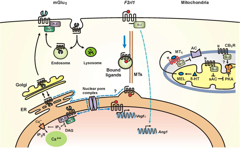
Fig. 1.
Schematic representation of various intracellular GPCRs. Left, proposed model of mGlu5 receptors trafficking in neurons in which >90% of mGlu5 traffics through the Golgi (27). Subsequently, 15–40% goes to the cell surface where it undergoes a cycle of constitutive endocytosis and recycling (27, 122). Alternatively, 60–85% of mGlu5 is retrogradely trafficked back to the endoplasmic reticulum (ER) and then, via lateral diffusion (dotted blue line), reaches the nuclear membrane (27, 123). Middle, ligand bound F2rll can translocate from the retinal ganglion cell surface to the nucleus along microtubules (MTs); nuclear F2rl1 activates vascular endothelial growth factor (Vegfα) expression. In contrast, signaling from cell surface F2rl1 results in the angiogenic gene, Angl expression (16). Right, a growing number of GPCRs have been localized to the outer (MT1, CB1 receptors) and inner mitochondrial membranes (AT2 receptor). Studies show that serotonin (5-HT) is converted into melatonin (MEL) within the mitochondrial matrix; MEL diffuses freely across membranes to activate MT1 receptors in the outer mitochondrial membrane (52). CB1 receptors have also been described in the outer mitochondrial membrane (124) although its downstream signaling machinery is primarily located within the matrix. The exact orientation of mitochondrial GPCRs and their signal transduction pathways is largely unknown. Other abbreviations include AC, adenylyl cyclase; sAC, soluble adenylyl cyclase; “I”, IP3R, inositol trisphosphate receptors; DAG, diacylglycerol; PLC, phospholipase C; Complex I; “P”, phosphorylation site on Complex I; black arrows indicate enhancement of activity; black bars indicate reduction of activity; αi inhibitory subunit of Gi protein; N, amino terminus; and PKA, protein kinase A.

