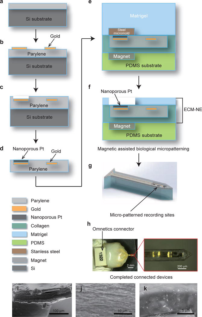
Figure 1.

Steps for fabricating ECM-based intracortical neural microelectrodes. (a)–(c) Steps for fabricating the ultra-thin parylene/conductor core; (d) electrochemical deposition of nanoporous Pt on the recording sites; (e)–(f) magnetic-assisted micropatterning of the top Matrigel layer; (g) illustration of the ECM-NEs after laser ablation. (h) A representative ECM-NE device and an enlarged view of the electrode tip. (i)–(k) SEM images of the cross-sectional areas of ECM-NEs: (i) the full device; (j) collagen; and (k) Matrigel.
