FIG.1.
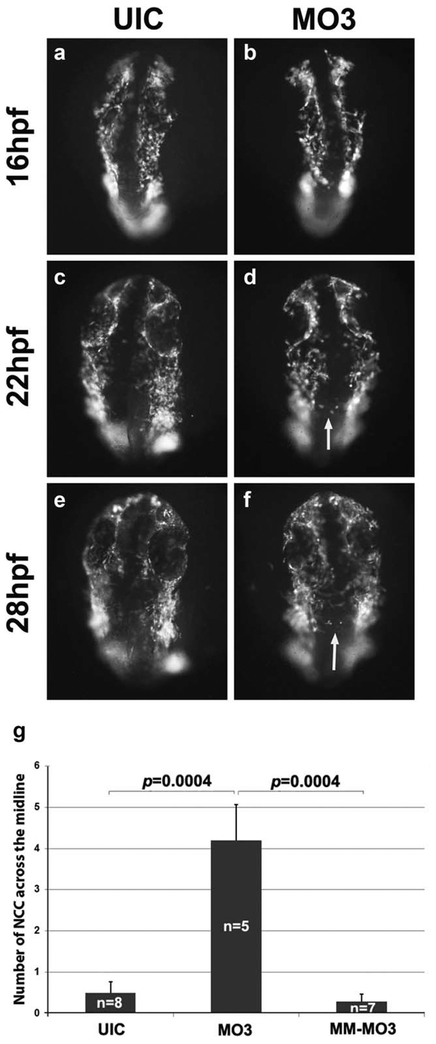
NCC migration is disorganized in MO3 morphants. Timelapse live cell imaging captures of sox10:GFP embryos showing migrating NCC cells at 16 (a, b), 22 (c, d), and 28 hpf (e, f). UIC (a, c, and e) and MO3-injected embryos (b, d, and f) showing a dorsal view of migrating NCCs. White arrows point to abnormally migrating NCCs. (g) Quantification of number of cells crossing the midline in UIC, MO3 and control MM-MO3 injected embryos.

