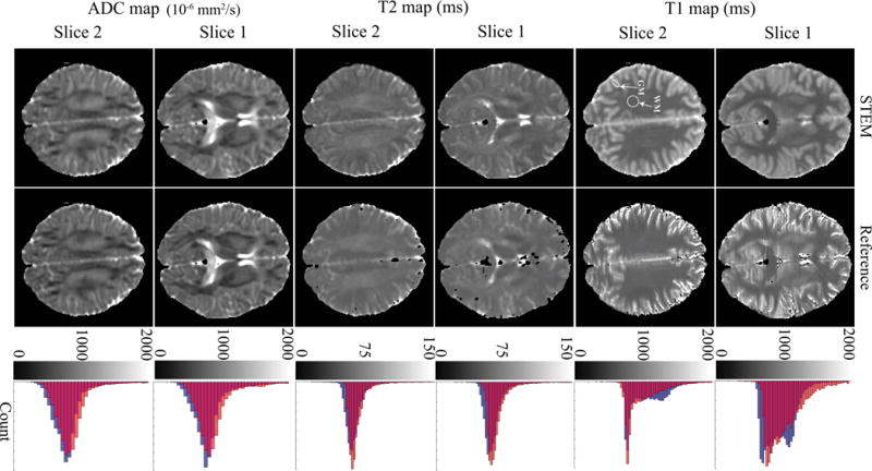
Figure 3.

The distributions of T1, T2 and ADC measurements in the brain from STEM are in good qualitative agreement with the reference measurements. The histograms to the right show the overall accuracy of each measurement from the entire slice. Orange bars in the histograms are from the reference maps, blue bars are measurements from STEM and the red color represents the overlapping area of reference and STEM-based histograms. The white arrows point to the ROIs in the white matter and gray matter.
